Apoptosis Signaling Pathway
Apoptosis is the process of programmed cell death characterized by distinct morphological characteristics and energy-dependent biochemical mechanisms. Apoptosis is considered a vital component of processes including normal cell turnover, proper development and functioning of the immune system, hormone-dependent atrophy, embryonic development and chemical-induced cell death.
Apoptotic cells undergo morphological changes involving extensive plasma membrane blebbing followed by karyorrhexis. Apoptotic bodies are formed by separation of cell fragments during a process called “budding.” Apoptotic bodies consist of cytoplasm with tightly packed organelles with or without a nuclear fragment. The organelle integrity remains enclosed within an intact plasma membrane. These bodies are subsequently phagocytosed by macrophages, parenchymal cells, or neoplastic cells and degraded within phagolysosomes. There are three main types of biochemical changes observed in apoptosis:
-
Activation of caspases,
-
Breakdown of DNA and protein and
-
Membrane changes and recognition by phagocytic cells.
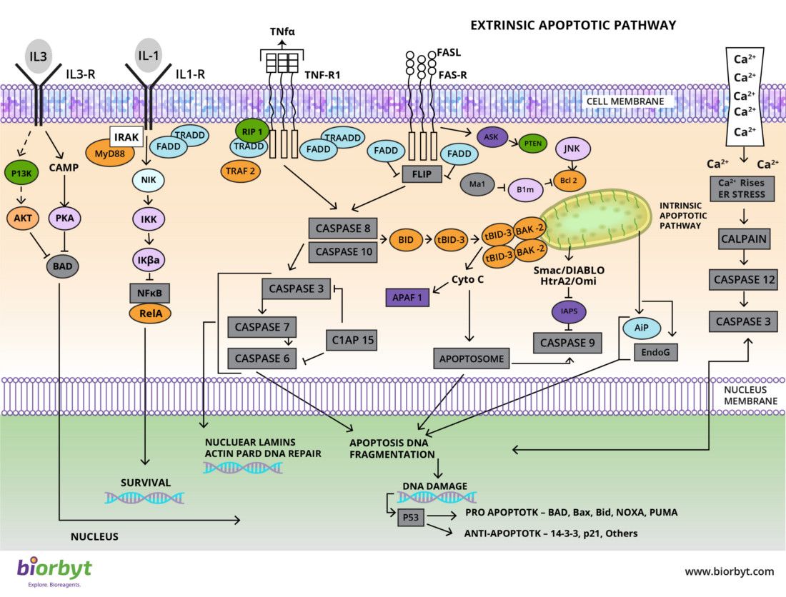
The two most widely characterised apoptotic pathways are the Intrinsic and Extrinsic pathways. The extrinsic signaling pathways involve transmembrane death receptors that are members of the tumor necrosis factor (TNF) receptor gene superfamily. Characterized ligands and corresponding death receptors include FasL/FasR, TNF-α/TNFR1, Apo3L/DR3, Apo2L/ DR4 and Apo2L/DR5. The intrinsic signaling pathways that initiate apoptosis are mitochondrial-initiated events involving a diverse array of non-receptor-mediated stimuli. These stimuli cause changes in the inner mitochondrial membrane that result in an opening of the mitochondrial permeability transition (MPT) pore, loss of the mitochondrial transmembrane potential and release of two main groups of normally sequestered pro-apoptotic proteins from the intermembrane space into the cytosol. These proteins activate the caspase- dependent mitochondrial pathway. Cytochrome C binds and activates Apaf-1 as well as procaspase-9, forming an “apoptosome”. The control and regulation of these apoptotic mitochondrial events occurs through members of the Bcl-2 family of proteins. The tumor suppressor protein p53 has a critical role in regulation of the Bcl-2 family of proteins.
Apoptosis Pathway Products – A Very Small Selection!
| Catalogue code | Description | Product Type |
|---|---|---|
| orb10173 | BCL2 | Antibody |
| orb183257 | DRAK2 | Antibody |
| orb385613 | CD95 | Antibody |
| orb10242 | Caspase 9 | Antibody |
| orb80955 | Human BCL2 | Protein |
| orb154650 | Cisplatin | Small Molecule |
| orb11024 | ASK1/MAPKKK5 | Antibody |
| orb419426 | Z-VAD-FMK | Small Molecule |
| orb50135 | Human Survivin | ELISA kit |
| orb223930 | Human TIGAR-TAT | Protein |
Apoptosis Pathway References
-
Ouyang, L. et al. Programmed cell death pathways in cancer: a review of apoptosis, autophagy and programmed necrosis. Cell Proliferation. (2012) 45, 487-498 doi: 10.1111/j.1365-2184.2012.00845.x
-
Ichim, G. and Tait, S. W. G. A fate worse than death: apoptosis as an oncogenic process. Nature Reviews Cancer. (2016) 16 539-548 doi:10.1038/nrc.2016.58
Helping your research
Biorbyt has an extensive range of small molecules available as research reagents to target proteins in the Apoptotic pathways. We provide the best service and a simple buying process allowing you to focus on your valuable research. Take a look at our product pages where you will find competitive pricing, lead times, and easy delivery options. If you have any questions, just ask us.
